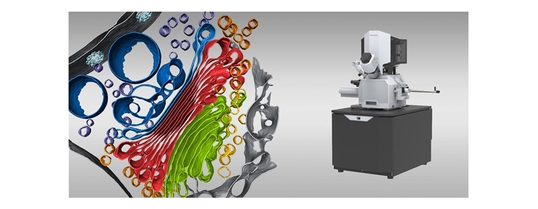
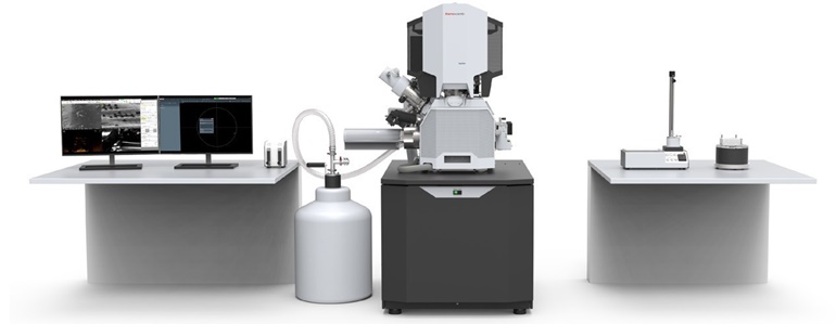
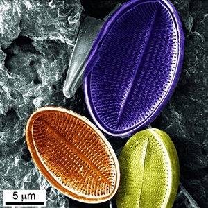
This is the first double-beam cryo microscope designed to capture a frozen, thin lamella from a biological sample.
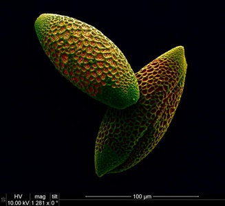 |
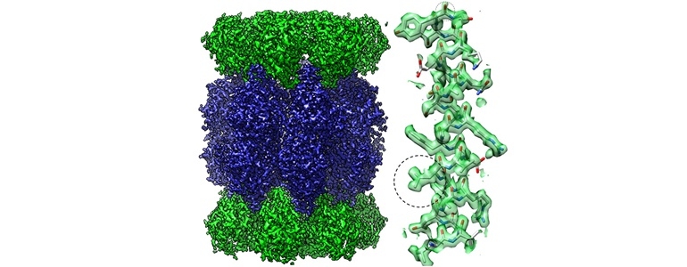
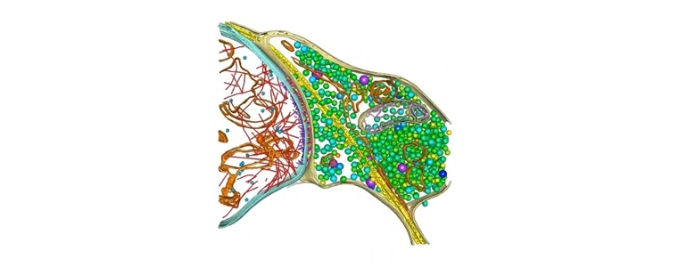
Cryo electron tomography is available to visualize the structure in its natural form. 3D visualization.
|
|
Automated processes using AutoTEM Software. The system works with ion focused plasma. This is a promising technique for anyone working in biology and medicine.
|
 |